Bayer Blood Sampling Kit
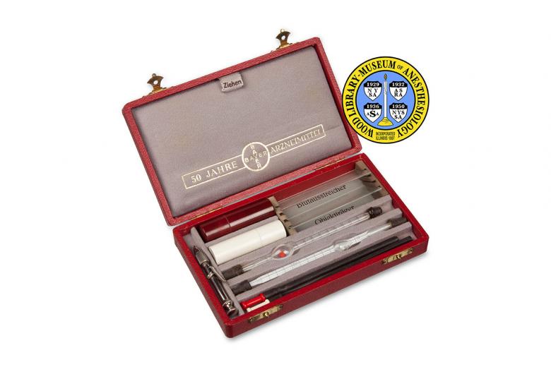
The blood sampling apparatus seen here dates from 1938, the fiftieth anniversary of Bayer's pharmaceutical division. In the early decades of the 20th Century, many hospitals had meager laboratory facilities, and rather than send blood to a laboratory, some anesthesiologists would have used kits like this one to conduct tests, such a red blood cell count, themselves.
The lancet in the kit was used to prick the patient’s ear lobe or finger to obtain a few drops of blood. One drop was tested using special blotting paper and a color scale, to determine the concentration of hemoglobin in the patient’s blood. Hemoglobin is the protein in red blood cells that carries oxygen. Another drop was smeared on a slide, and some drops were collected with the rubber suction tubes and glass pipettes to be mixed with a small amount of one of the solutions contained in the vials (one for red blood cells and one for white blood cells). The carefully diluted samples would be placed on specially constructed slides for examination under a microscope. These tests could then help the anesthesiologist determine whether there were any problems that required treatment, such as dehydration or anemia.
Catalog Record: Bayer Blood Sampling Kit
Access Key: akhl
Accession No.: 2012-12-21-2
Title: Blutentnahme-Besteck für hämatologische Untersuchungen / Bayer.
Corporate Author: Bayer.
Title variation: Alt Title
Title: Blood sampling kit.
Title variation: Alt Title
Title: Blood cutlery for haematological investigations.
Publisher: Germany : Bayer, [1938?].
Physical Descript: 1 hematologic kit : wood, glass, metals, rubber, plastic, textiles ; 3.5 x 16
x 10 cm.
Subject: Hematologic Tests – instrumentation.
Subject: Erythrocyte Count – instrumentation.
Note Type: General
Notes: Date based on the marking on the inside of the box that announces the 50th
anniversary of Bayer Pharmaceuticals: “50 JAHRE BAYER ARZNEIMITTEL”.
Note Type: Citation
Notes: Berger D. A brief history of medical diagnosis and the birth of the clinical
laboratory: Part 2 – laboratory science and professional certification in the
20th century. MLO Med Lab Obs. 1999;31(8):32-34,36,38.
Note Type: Citation
Notes: Das Blut und seine Zusammensetzung (eine kurze Übersicht). Germany: Bayer;
[1938].
Note Type: Citation
Notes: Burke MD. Laboratory medicine in the 21st century. Am J Clin Pathol.
2000;114(6):841-846.
Note Type: Citation
Notes: Kotlarz WR. Tracing our roots: origins of clinical laboratory science. Clin
Lab Sci. 1998;11(1):5-7.
Note Type: Citation
Notes: Ochei J, Kolhatkar A. Medical Laboratory Science: Theory and Practice. New
Delhi: Tata McGraw-Hill; 2000:277-278.
Note Type: Citation
Notes: Ochsner AJ, Percy NM. General surgical considerations. A New Manual of
Surgery: Civil and Military. 6th ed. Chicago: Cleveland Press; 1917:40-42.
Note Type: Citation
Notes: Race GJ, Tillery GW, Dysert PA 2nd. A history of pathology and laboratory
medicine at Baylor University Medical Center. Proc (Bayl Univ Med Cent). 2004
Jan;17(1):42-55.
Note Type: Physical Description
Notes: A wood[?] box covered in deep red buckram or leather measuring 3.1 cm in
height, 15.8 cm in width and 9.7 cm in depth (including latches and hinges);
Two brass colored hinges hold the box closed in front; Marked in gold
lettering on the top of the box is, “Blutentnahme-Besteck [new line] für [new
line] hämatologische Untersuchungen”; Imprinted on the bottom of the box is,
“Made in Germany”; The bottom of the inside of the box is divided into slots
to hold the instruments and supplies in the kit; The instruments include a
stainless steel lancet, two ruber tubes (one with a red mouthpiece and one
with a white mouthpiece), one red cell Thoma-style mixing pipette, one white
cell Thoma-style mixing pipette, and two glass slides; There is also one red
plastic vial with a screw-on top that holds one glass vial with a black
rubber stopper; This glass vial once contained diluting liquid for red blood
cells (Hayemsche solution); There is also a white plastic vial with a
screw-on top that holds one glass vial with a black rubber stopper; This
glass vial once contained diluting solution for white blood cells; Printed on
the inside of the bottom of the box below the area for the glass slides are
the words, “Blutausstreicher” and “Objektträger”; Inside the lid of the box
is a cushion covered with light grey fabric that is marked with gold colored
print, “50 JAHRE BAYER ARZNEIMITTEL”; Pulling on a fabric tab attached to one
edge of the cushion, and marked, “Ziehen”, reveals a booklet stored behind
the cushion; The cover title of the booklet is, “Das Blut und seine
Zusammensetzung (eine kurze Übersicht)”; On the first page of the booklet is
an advertisement for the drug, “CAMPOLON”; A photograph at the end of the
booklet shows packaging for CAMPOLON; Other pages in the booklet (all written
in German) provide instruction for using the kit to make a blood smear, and
prepare a dilution of red and white blood cells for manual cell counts
(hemocytometer not included); Reference tables for blood and its composition
and morphology, and a Haemoglobin color scale with blotting paper and
instructions; A sticker on the back of the booklet is printed with the
following text, “50 JAHRE [new line] Arzneimittel [new line] mit dem [Bayer
logo] Bayer-Kreuz [new line] Das Zeichen Des Vertrauens”.
Note Type: Reproduction
Notes: Photographed by Mr. Steve Donisch on January 15, 2013.
Note Type: Historical
Notes: During the first few decades of the 20th century many US hospitals had meager laboratory facilities. Often the lab consisted of little more than a microscope, a few reagent bottles, and some test tubes and slides, which were poorly maintained and located in the hospital’s basement or a small corner in a ward. As more laboratory tests became available and methods improved, facilities and staffing for clinical laboratories expanded. In 1923, 48% of U.S. hospitals had in-house clinical laboratories. Growth of clinical laboratories occurred slowly for some hospitals especially up to World War II. During this time, if an anesthesiologist wanted a hemoglobin or red blood cell count for a patient, depending on the urgency, the staffing and the funding of the laboratory at his (or her) hospital, he may have performed the test himself using tools very much like those contained in this kit.
Note Type: Historical
Notes: This kit was released by the German pharmaceutical company Bayer in 1938 as part of a marketing campaign for their new medication for the treatment of anemia, called Campolon, and in recognition of the company’s fiftieth anniversary. It came with an instruction booklet, blotting paper, and a hemoglobin color scale.
Note Type: Historical
Notes: This pocket kit contains the tools that a physician would have needed in order to obtain a few drops of blood and prepare them for four or so basic tests. The lancet, located on the left side of the kit, includes a thick, short needle that was used to prick the skin of the ear lobe or finger. A drop of blood was placed on a special kind of blotting paper that was then compared with a color scale that came with the kit. The color on the scale that most closely matched the color of the blood on the paper determined the estimate of the patient’s hemoglobin concentration. Hemoglobin is the protein in red blood cells that carries oxygen, and it is one laboratory result considered when diagnosing anemia.
Next the physician would use the thin black suction tubes, one with a red mouthpiece and one with a white
mouthpiece, to attach to the corresponding Thoma pipette (the glass pipettes with a central bulb containing a bead) and carefully suction a small amount of another drop of blood to a marked line. The physician would then use the
corresponding vials (the red vial contained a solution for diluting red blood cells, and the white vial contained a solution for diluting white blood cells) to slowly suction the proper amount of solution into the pipette with
the blood. The pipettes would be twirled until the blood and solutions were thoroughly mixed, and then capped to hold the dilution within the pipettes until the physician could get to a laboratory with a microscope and
hemocytometer (a special kind of slide and cover that aids in the counting of the very small blood cells).
Finally, the physician would use the slides in the kit to allow a final drop of blood to fall on one end of a slide. The
drop was then smeared, with the other slide, and allowed to air-dry. Later, the physician would apply a stain to the dried blood smear to exam it under the microscope.
Note Type: Exhibition
Notes: Selected for the WLM website.
