Flagg Haemoxometer
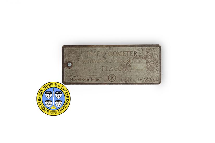
During surgery, anesthesiologists monitor and manage changes in their patients’ vital functions, including breathing and blood oxygenation. This device, a haemoxometer or oxyhaemoglobinometer, was designed by Dr. Paulel J. Flagg, (1886-1970) as a means of estimating how much oxygen was circulating in a patient’s blood.
When Dr. Flagg designed this tool, anesthesiologists were already monitoring their patients’ skin color. Skin color is an important part of any medical assessment, but it is also an imprecise way to gauge oxygen levels. Dr. Flagg believed his haemoxometer would greatly improve anesthesiologists’ ability to accurately estimate their patients’ oxygen levels. He also thought its use would enable anesthesiologists to intervene in a more timely manner.
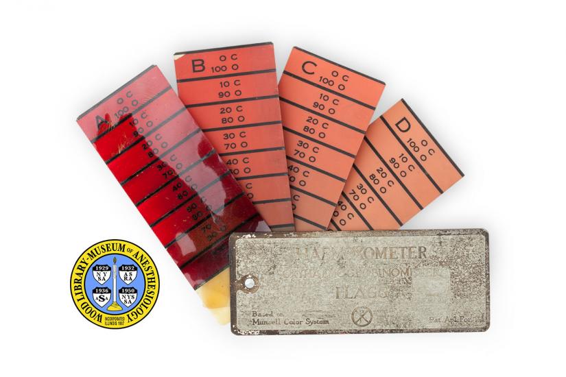
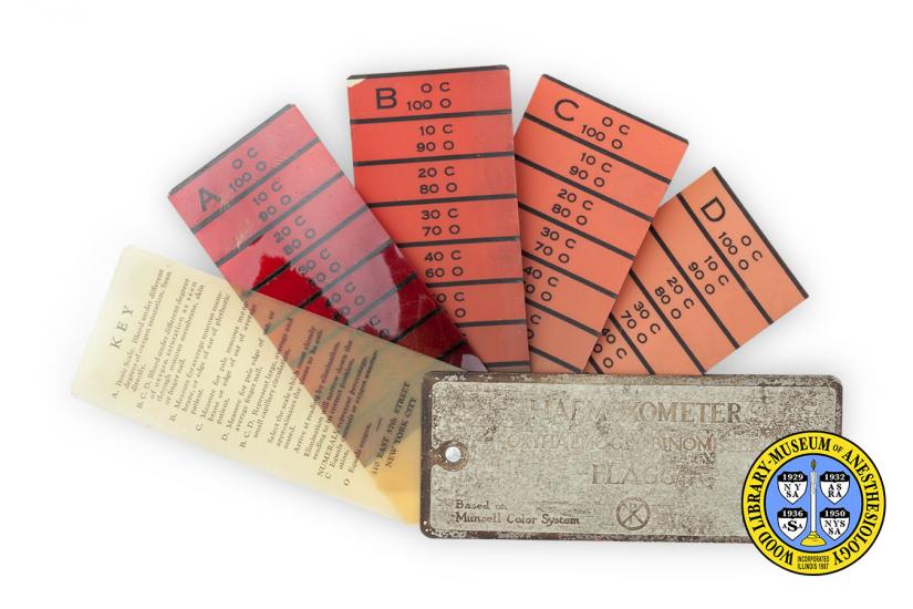
Dr. Flagg worked with a color expert familiar with the Munsell System, which represents colors according to three attributes: hue, value and chroma. They constructed a series of scales that represent various blood oxygen saturations as seen in the color of the nails, skin and mucous membranes of patients under anesthesia. Color scales where not included for people with dark skin tones or for those with conditions that significantly change skin tone, like jaundice. For these circumstances Dr. Flagg recommended using the color of the tissue and blood at the surgical site.
Catalog Record: Flagg Haemoxmeter
Access Key: akwt
Accession No.: 96
Title: Haemoxometer oxyhaemoglobinometer / Flagg.
Author: Flagg, Paluel J. (Paluel Joseph), 1886-1970.
Title variation: Alt Title
Title: Flagg haemoxometer oxyhaemoglobinometer.
Title variation: Alt Title
Title: P. J. Flagg’s haemoxometer.
Title variation: Alt Title
Title: Hemoxometer.
Publisher: New York : [Maryknoll?], [between 1922 and 1924].
Physical Descript: 1 monitoring device : metal and plastics ; 1 x 13 x 5.5 cm.
Subject: Monitoring, Intraoperative – instrumentation.
Subject: Oxygen.
Subject: Cyanosis – prevention and control.
Note Type: General
Notes: The early year in the date range for the possible year of manufacture is
based on the year that Dr. Flagg applied to patent the device and first
described it in a publication. The end-date in the date range is an estimate
based on the year that the device was patented (1924). This item was included
in a 1938 inventory by Dr. Wood.
Note Type: General
Notes: The address marked on the object (410 East 57th Street) was the address for
the Maryknoll Medical Mission during the possible years of production.
Note Type: Citation
Notes: Bause GS. P. J. Flagg’s haemoxometer.
Note Type: Citation
Notes: Dr. Paluel Joseph Flagg. Catholic Medical Mission Board website. https://cmmb.
org/dr-paluel-joseph-flagg. Accessed April 3, 2013.
Note Type: Citation
Notes: Flagg PJ, inventor. Method and apparatus for determining the amount of oxygen
combined with the haemoglobin of blood. US patent 1,513,542. October 28, 1924
Note Type: Citation
Notes: Flagg PJ. An oxyhaemoglobinometer for the clinical measurement of cyanosis.
Proc Soc Exp Biol Med. 1922;20(1):1-5.
Note Type: Citation
Notes: Flagg PJ. The signs of anaesthesia. The Art of Anaesthesia. 3rd ed.
Philadelphia: J.B. Lippincott Company; 1944:96-102.
Note Type: Citation
Notes: Flagg PJ. The signs of anaesthesia. The Art of Anaesthesia. 7th ed.
Philadelphia: J.B. Lippincott Company; 1944:96-102.
Note Type: Citation
Notes: Sister Virginia Flagg, MM. Maryknoll Mission Archives.
Accessed April 3, 2013.
Note Type: Citation
Notes: Wiest JP. A history of mission in China. In: Kroeger JH, ed. The Gift of
Mission: Yesterday, Today, Tomorrow – Maryknoll Centennial Symposium. New
York: Orbis Books; 2013:87-88.
Note Type: Physical Description
Notes: One hemoxometer composed of two rectangular metal plates hinged together at
one end, with five plastic index cards between them; Four of the cards are
labeled with a different capital letter, A, B, C and D; Each of these four
cards are also marked with eight areas of different saturations of red,
marked from top to bottom with black lines between each area: “O C [new line]
100 O”, “10 C [new line] 90 O”, “20 C [new line] 80 O”, “30 C [new line] 70
O”, “40 C [new line] 60 O”, “50 C [new line] 50 O”, and “70 C [new line] 30
O”; The colors have faded and changed with time; One of the plastic cards is
a transparent yellow and marked at the top with, “KEY”; The remainder of the
text on the key includes, “A. Basic Scale. Blood under different degrees of
oxygen saturation. Seen directly. [new line] B. C. D. Blood under different
degrees of oxygen saturation as seen through mucous membrane, skin or finger
nail. [new line] B. Measure for average mucous membrane, or edge of ear of
plethoric patient. [new line] C. Measure for pale mucous membrane or edge of
ear of average patient. [new line] D. Measure for pale edge of ear, or
average finger nail. [new line] B.C. D. Represent large, average and small
capillary circulation. [new line] Select the scale which most closely
approximates the tissue to be estimated.[new line] Arrive at a reading by
elimination. Elimination will narrow down the reading to its correct position
[new line] NUMERALS represent percentage. [new line] C equals cyanosis or
oxygen unsaturation. [new line] O equals oxygen. [new line] 410 EAST 57th
STREET [new line] NEW YORK CITY”; The top metal plate has the following
markings: “HAEMOXOMETER [new line] OXYHAEMOGLOBINOMETER [new line] FLAGG [new
line] Based on Munsell Color System [Chi Rho symbol] Pat. Apl. For”.
Note Type: Reproduction
Notes: Photographed by Mr. Steve Donisch, September 18, 2013.
Note Type: Historical
Notes: During surgery, anesthesiologists monitor and manage changes in their
patients’ vital functions, including breathing and oxygenation. This device,
a haemoxometer or oxyhaemoglobinometer, was designed by Dr. Paulel J. Flagg,
(1886-1970) as a means of estimating how much oxygen was circulating in a
patient’s blood.
When Dr. Flagg designed this tool, anesthesiologists were already monitoring
their patients’ skin color. Skin color is an important part of any medical
assessment, but it is also an imprecise way to gauge oxygen levels.
Dr. Flagg believed his haemoxometer would greatly improve anesthesiologists’
ability to accurately estimate their patients’ oxygen levels. He also thought
its use would enable anesthesiologists to intervene in a more timely manner.
Dr. Flagg worked with a color expert familiar with the Munsell System, which
represents colors according to three attributes: hue, value and chroma. They
constructed a series of scales that represent various blood oxygen
saturations as seen in the color of the nails, skin and mucous membranes of
patients under anesthesia. Color scales where not included for people with
dark skin tones or for those with conditions that significantly change skin
tone, like jaundice. For these circumstances Dr. Flagg recommended using the
color of the tissue and blood at the surgical site.
Flagg’s Haemoxometer was introduced in 1922, about 60 years before the
introduction of pulse-oximeters. The Manufacturer of this haemoxometer was
Maryknoll. Paulel Flagg was a representative for the Maryknoll Medical
Mission located in New York City. Maryknoll was a supporting organization of
the Catholic Medical Mission Board, established in 1924 by a group of
volunteers that included Dr. Flagg. Dr. Flagg’s daughter, Sister Virginia
Flagg (1912-2011), joined Maryknoll Missions in 1930, and served with them
for 64 years before she retired.
Note Type: Exhibition
Notes: Selected for the WLM website.
