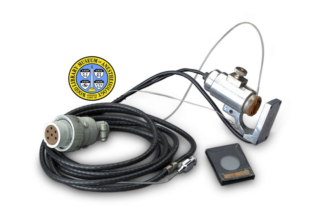
Waters X-350 Ear Oximeter
Anesthesiologists monitor and manage the surgical patient’s breathing and other vital functions. Beginning in the 1940s, the increasing use of mechanical ventilators in anesthesia led to a demand for better oxygen monitors. An oximeter continuously monitors the amount of oxygen in a person’s blood. By sending two or more wavelengths of light through the thinnest parts of the body, such as the ear, and then reading the extent to which that light is absorbed by the blood and tissues, transmission oximeters provided an accurate, noninvasive measurement of blood oxygenation.
The first ear oximeter was introduced in 1942 by American physiologist, Glenn A. Milliken (1906-1947), for use in military aviation. In 1949, it was modified by Earl H. Wood, M.D. (1912-2009), and his colleagues at the Mayo Clinic, in Rochester, MN, who were also working on problems of military aviation. Dr. Wood’s oximeter was made by Waters Conley Co. of Minneapolis, MN.
The successor to that firm, Waters Company of Rochester, MN, went on to make commercial oximeters and other medical equipment. The earpiece shown here is one component of the Waters X-350 ear oximeter, advertised in the 1960s. The screw could be loosened to open and slide the unit over the rim of the patient’s ear (the pinna), then tightened to hold it in place. An improved technology, pulse oximetry, became widely available in the 1980s.
Catalog Record: Waters X-350 Ear Oximeter Waters X-350 Ear Oximeter
Access Key: andv
Accession No.: 2012-05-30-1 D
Title: [ X-350 oximeter earpiece / Waters Company].
Corporate Author: Waters Corporation.
Publisher: Rochester, Minnesota : The Waters Company, [between 1954 and 1983].
Physical Description: 1 oximeter earpiece : metals, plastics, rubber ; 3.5 x 10 x 227.5 cm.
Subject: Blood Gas Monitoring, Transcutaneous – instrumentation.
Subject: Oxygen – analysis.
Subject: Oximetry – instrumentation.
Web Link: https://www.woodlibrarymuseum.org/museum/item/1028/waters-x-350-ear-oximeter
Note Type: General
Notes: The title is taken from product literature in the Waters Company file. The first year in the date range is the year that Mr. George Waters left Waters Conley Co. and founded The Waters Company. The second year in the date range is the year that the Nellcor 100 pulse oximeter was introduced. This description orients the earpiece with the front (opening) side facing (proximal to) the patient, and the tubes extending from the back (distal to the patient.)
Note Type: Citation
Notes: American Society of Anesthesiologists. Standards for Basic Intra-Operative Monitoring. Park Ridge, Illinois: American Society of Anesthesiologists, 1989. Standards, Guidelines and Statements Series. American Society of Anesthesiologists Archives. Located at: Wood Library-Museum of Anesthesiology, Schaumburg, Illinois.
Note Type: Citation
Notes: Earl H. (Howard) Wood. Minnesota Science and Technology Hall of Fame website. http://www.msthalloffame.org/earl_wood.htm. Accessed June 21, 2016.
Note Type: Citation
Notes: All Nobel Prizes in Physics. Nobel Foundation website. http://www.nobelprize.org/nobel_prizes/physics/laureates/. Accessed June 21, 2016.
Note Type: Citation
Notes: Payne JP, Severinghaus JW, eds. Pulse Oximetry. London: Springer-Verlag, 1986.
Note Type: Citation
Notes: A Historical Perspective. RT Magazine website. http://www.rtmagazine.com/2007/02/a-historical-perspective/. Accessed June 20, 2016.
Note Type: Citation
Notes: Severinghaus JW, Astrup PB. History of Blood Gas Analysis. Boston: Little, Brown, 1987. International Anesthesiology Clinics. Winter, 1987;24(4):167-204.
Note Type: Citation
Notes: George Franklin Waters [Obituary]. Star Tribune Media Company website. http://www.startribune.com/obituaries/detail/85646/?fullname=george-franklin-waters. Accessed June 16, 2016.
Note Type: Citation
Notes: Waters Company [company file]. Archives. Located at: Wood Library-Museum of Anesthesiology, Schaumburg, Illinois.
Note Type: Physical Description
Notes: One oximeter earpiece; The metal head measures 1.5 x 4 x 3.5 centimeters; From front to back, the head has an oblong shape that is wider on the proximal side; The head is comprised of two halves that can be adjusted to stand either together or apart; When completely closed, the upper and lower halves of the head meet, and the head resembles a squared capital letter O; When open, the head resembles a squared capital letter G; The open ends of the G are fitted with plastic discs; The disc on the upper half of the G is held in place by two screws; The disc on the lower half of the G is held in place by both a metal ring and a black rubber ring;
The broad, top side of the head has the number “466” incised in the upper half; In this orientation, the number is upside-down; The top side of the lower half is featureless; The bottom side of the head holds four wires, one yellow, one blue, one green and one white; In the upper half on this side, each wire enters one of a vertical row of four small holes; In the lower half, all four wires enter a single hole; When held between index finger and thumb, the top edge of the head has a striated surface, over which the flat cap of the post is fitted at the distal end, while the bottom edge is smooth and holds two small screws;
A rod is seated in the distal side of the lower half; a corresponding tube on the distal side of the upper half can slide up and down over that rod; Extending the upper half in this way creates an opening on the proximal side; When the upper half is fully extended, the opening is 1 centimeter high; The distal side of the upper half holds a set screw that can be loosened to allow adjustment of the two halves, and tightened to hold the two halves in the desired position;
Two tubes enter the distal side of the lower half of the metal head; The upper of these two tubes is made of clear plastic and the lower is made of black plastic; These two tubes are lashed together by four cut sections of black plastic tubing; The black plastic tube is approximately 167 centimeters long; The opposite end of the black plastic tube is attached to an electrical fitting that holds a rosette of six prongs;
The clear plastic tube is approximately 44.5 centimeters long; The opposite end of this tube is fitted over a needle; The hub of the needle is marked “B-D [new line] 20”’; The hub of the needle is fixed in a metal connector; The same connector has a side clamp that grips the black plastic tube; At the end that is opposite to the needle, this connector is fitted to a long black rubber tube; This rubber tube is marked “Conductive”, it is approximately .5 centimeter in diameter, and is approximately 86.5 centimeters long;
At the end that is opposite to the connector which holds the needle, the long rubber tube is attached to one leg of a translucent white plastic Y connector; The opposite end of the Y connector is attached to a second, short piece of black rubber tube, approximately 13.5 centimeters long; This second rubber tube is attached at its opposite end to a metal fitting that holds a black rubber hand-bulb; This metal fitting is marked “AIR-FLO [new line] CONTROL” [new line] “W. A. BAUM CO. [new line] COPIAGUE NY”; The second leg of the Y connector is attached to a third piece of black rubber tubing, approximately 11.5 centimeters long, which ends in a metal fitting.
Note Type: Reproduction
Notes: Photographed by Mr. Steve Donisch, January 13, 2016.
Note Type: Acquisition
Notes: Gift of Diane Bohlman, M.D.
Note Type: Historical
Notes: Anesthesiologists monitor and manage the surgical patient’s breathing and other vital functions. The introduction of muscle relaxant drugs in the 1940s, and the polio epidemics of the 1950s, led to an increasing use of mechanical ventilators in anesthesia. These developments, together with improvements in heart surgery and the needs of high-altitude aviation, created a demand for better oxygen monitors.
The role of hemoglobin as the carrier of oxygen in the blood was first understood in the 1860s. In 1874, German physician Karl von Vierordt (1818-1884) studied the oxygenation of his own blood by using a spectroscope to measure light that was passed through his hand. In the 1930s, the use of photoelectric cells allowed more accurate measurement of blood gases, and the first oximeters were created.
An oximeter continuously monitors the amount of oxygen in a person’s blood. By sending two or more wavelengths of light through the thinnest parts of the body, such as the ear, and then reading the extent to which that light is absorbed by the blood and tissues, the devices called transmission oximeters provide an accurate, noninvasive measurement of the oxygen saturation of hemoglobin in arterial blood. The first ear oximeter was introduced in 1942 by American physiologist, Glenn A. Milliken (1906-1947), who also coined the word “oximetry”. Developed for use in military aviation, the device used mercury vapor light, colored filters and a photoelectric cell. Milliken’s oximeter was modified in 1949 by Earl H. Wood, M.D. (1912-2009) and his colleagues at the Mayo Clinic, in Rochester, MN. This team, also, were working on problems of military aviation.
Dr. Wood’s oximeter was made by Waters Conley Co. of Minneapolis, MN. The successor to that firm, The Waters Company of Rochester, MN, went on to make commercial oximeters and other medical equipment. The earpiece described in this record is just one component of a Waters X-350 ear oximeter, which was advertised in the 1960s. The screw could be loosened to open and slide the unit over the rim of the patient’s ear (the pinna), then tightened to hold it in place. An improved technology, pulse oximetry, became widely available in the 1980s. In 1989, the American Society of Anesthesiologists adopted new Standards for Basic Intra-Operative Monitoring, requiring assessment of the patient’s blood oxygenation during all anesthetics.
Note Type: Exhibition
Notes: Selected for the WLM website.
