Arndt Endobronchial Blocker
Surgeries that involve a lung require special anesthesia techniques to ensure that patients continue to breathe effectively. One method involves blocking airflow to the lung on the side of the chest being operated on, while continuing to ventilate the lung on the other side of the chest.
The technique of blocking airflow to a lung or section of a lung is called bronchial blockade. It involves the insertion of a catheter or tube down the trachea (windpipe) and into one of the main bronchi (primary tubes to the lungs), or into one of the smaller bronchi that ventilate just a section of a lung. A balloon at the end of the blocker’s tube is then inflated to seal off the lung. The first endobronchial blocker was introduced around 1935.
In 1997, anesthesiologist George A. Arndt, M.D., of Madison, Wisconsin, introduced a new design that made it easier to correctly insert an endobronchial blocker into position. Dr. Arndt’s blocker was a thin catheter with an inner wire that was looped at the very end. The loop was used to connect the catheter to a flexible fiberoptic bronchoscope. This allowed video imaging to be used to place the catheter through a normal endotracheal (breathing) tube. Dr. Arndt also designed an accompanying multiport adapter that allowed the patient to be ventilated while the blocker was being placed. Also, the balloon on Dr. Arndt’s catheter exerted less pressure against the delicate tissue inside the bronchi than did most other balloon-tipped catheters at that time.
Catalog Record: Arndt Endobronchial Blocker
Access Key: amqt
Accession No.: 2005-06-11-1 C
Title: [Wire-guided endobronchial blocker] / [George A. Arndt, Frank J. Fischer, Jr.].
Author: Arndt, George A.
Author: Fischer, Frank J.
Corporate Author: Cook Incorporated.
Title variation: Alt Title
Title: Arndt endobronchial blocker.
Title variation: Alt Title
Title: Arndt bronchial blocker.
Publisher: [Bloomington, Indiana] : Cook, [between 1997 and 2005?].
Physical Descript: 1 bronchial blocker : plastics, wire ; 93 x 20 x 2.5 cm.
Subject: Anesthesia, Thoracic – instrumentation.
Subject: One-Lung Ventilation – instrumentation.
Note Type: General
Notes: The early year in the date range for the possible year of manufacture is
based on the year that Arndt, Fisher and Cook Incorporated filled to patent
the catheter (1997). The end year is based on the year that the catheter was
donated to the WLM 2005). The date range could change if documentation or
expert opinion indicate the range should be corrected.
Note Type: General
Notes: The title is based on the name most commonly used to refer to the catheter in
the titles and abstracts of four 1999 publications that mention the invention
This includes the patent and the three 1999 articles recorded as citations.
Note Type: Citation
Notes: Arndt GA, Buchika S, Kranner PW, DeLessio ST. Wire-guided endobronchial
blockade in a patient with a limited mouth opening. Can J Anesth.
1999;46(1):87-89. https://link.springer.com/article/10.1007%2FBF03012521.
Accessed October 29, 2015.
Note Type: Citation
Notes: Arndt GA, DeLessio S, Kranner P. A new method to achieve one-lung ventilation
using a fiberoptically directable endobronchial blocker [abstract P103]. Acta
Anaesthesiol Scand. 1997;41(S110):188.
Note Type: Citation
Notes: Arndt GA, DeLessio ST, Kranner PW, Orzpowski W, Ceranski B, Valtysson B.
One-lung ventilation when intubation is difficult – presentation of a new
endobronchial blocker. Acta Anaesthesiol Scand. 1999;43(3):356-358.
https://onlinelibrary.wiley.com/doi/10.1034/j.1399-6576.1999.430320.
x/abstract;jsessionid=1ED496CA8436E9920598CE84621D77E5.f01t01. Accessed
October 29, 2015.
Note Type: Citation
Notes: Arndt GA, Fischer J Jr., inventors. Cook Incorporated, assignee. Guided
endobronchial blocker catheter. U.S. patent 5,904,648. May 18, 1999. www.
google.com/patents/US5904648. Accessed October 29, 2015.
Note Type: Citation
Notes: Arndt G. Wire-guided endobronchial blockage: an alternative means for
achieving one-lung ventilation. Internet J Anesthesiol. 1999;4(1).
https://ispub.com/IJA/4/1/3257. Accessed October 29, 2015.
Note Type: Citation
Notes: Brodsky JB. The evolution of thoracic anesthesia. Thorac Surg Clin.
2005;15(1):1-10.
Note Type: Citation
Notes: Brodsky JB, Lemmens HJ. The history of anesthesia for thoracic surgery.
Minerva Anestesiol. 2007 Oct;73(10):513-524. https://www.minervamedica.
it/en/journals/minerva-anestesiologica/article.php?cod=R02Y2007N10A0513.
Accessed October 30, 2015.
Note Type: Citation
Notes: Campbell L, Katz JA. Thoracic anestesia. In: Sikka PK, Beaman ST, Street JA,
eds. Basic Clinical Anesthesia. New York: Springer; 2015:384-385.
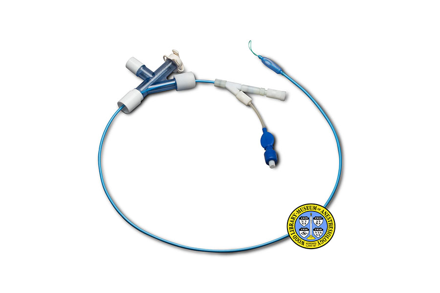
Note Type: Physical Description
Notes: One wire guided catheter for endobronchial blockade; Measurements were based
on the photograph taken by Mr. Donisch in June; The side facing up was
treated as the ‘front’ of the catheter; The length was treated as the
‘height’ and was taken with the catheter held in a linear position; The
distal end (from the patient’s perspective) and the extension for the pilot
balloon were allowed to lay in a ‘natural’ position for the measurements; The
catheter is a blue plastic with an expandable balloon at the proximal (from
the patient’s perspective) end; The guidewire is a light green and can be
removed from the catheter at the distal end; The port for inflating the
catheter balloon is located at the end of a side extension at the distal end
of the catheter; The catheter was photographed with the accompanying ‘special
bronchoscopy port’, also called the Cook Multiport Ventilating Adapter; The
multiport adapter is made of a clear blue plastic with white plastic port
ends, with the exception of the cap of the bronchoscope port, which is made
of white silicone rubber[?]; In the photograph the catheter is shown threaded
through the multiport adapter side extension made for it; The ventilation
port extends from the multiport adapter at a perpendicular angle; The
attachment for a bronchoscope is the most distal port; The proximal end of
the multiport adapter is for the attachment for the distal end of an
endotracheal tube; The only manufacturer markings are slightly elevated
lettering on both sides of the multiport adapter, “COOK”.
Note Type: Reproduction
Notes: Photographed for the WLM by Mr. Steve Donisch in June, 2015.
Note Type: Acquisition
Notes: Donated to the WLM by George Arndt, M.D. Donated facilitated by Mark E.
Schroeder, M.D.
Note Type: Historical
Notes: Surgeries that involve a lung require special anesthesia techniques to ensure
that effective ventilation and respiration continue to occur despite the
exposure of a lung to atmospheric pressure. One method involves blocking
airflow to the lung on the side of the chest being operated on, while
continuing to ventilate the lung on the other side of the chest.
The technique of blocking airflow to a lung or section of a lung is called
bronchial blockade. It involves the use of a tube or catheter with an
inflatable balloon at one end. The balloon is inflated after the tube has
been carefully inserted down the trachea and into one of the main bronchi, or
one of the smaller bronchi that ventilate just a section of a lung. The
inflation of the balloon seals off the bronchus and prevents air from moving
any further into the lung. The first bronchial blocker was introduced around
1935 by Canadian surgeon Edward W. Archibald (1872-1945).
In 1997, anesthesiologist George A. Arndt, M.D., of Madison, Wisconsin,
introduced a new design that made it easier to insert an endobronchial
blocker into the correct position. Dr. Arndt’s blocker was a thin catheter
with an inner wire that had a loop at the very end. The loop was used to
connect the catheter to a flexible fiberoptic bronchoscope. This allowed
video imaging to be used to place the catheter through a normal endotracheal
(breathing) tube. Dr. Arndt also designed an accompanying multiport adapter
that allowed the patient to be ventilated while the blocker was being placed.
Also, the balloon on Dr. Arndt’s catheter exerted less pressure against the
inside of the bronchus than did other balloon-tipped catheters at that time.
This was much better for the delicate tissue inside the airway.
Note Type: Exhibition
Notes: Selected for the WLM website (noted October 29, 205).
