Godart Capnometer
Anesthesiologists use capnographs to monitor how much carbon dioxide (CO2) is in a patient’s exhaled breath. They provide numeric values and a waveform of the increase and decrease in CO2 as the patient breathes. This wave form is called a capnogram. Anesthesiologists use the data to detect problems with the patient's condition, as well as problems with equipment.
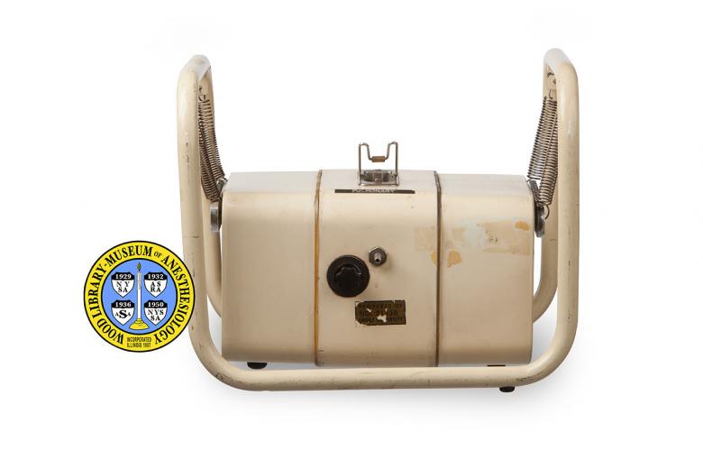
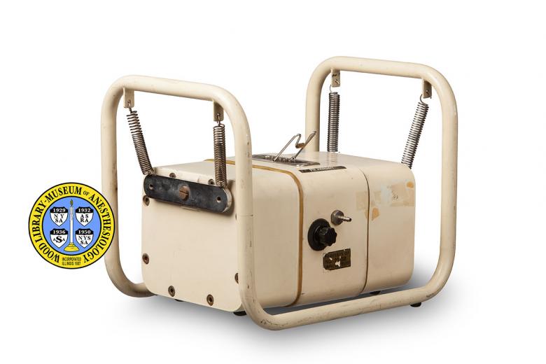
Most capnographs measure CO2 via infrared CO2 detectors called Luft Detectors, which were first introduced by Karl Friedrich Luft (1909-1999) in 1943. Use of the technology in the medical setting began in the 1950s. Because the data from the detectors has to be amplified, calculated, and converted into signals for display and graphing, early capnographs were large multi-piece machines. The Godart detector-head, or analyzer, pictured here was the smallest part of the Godart Capnograph. The patient's exhaled air was pumped from the breathing tube to the detector-head where the infrared Luft detectors measured the CO2. Via a cord plugged into the socket on the top of the detector-head, data was sent to the piece of the capnograph that housed the computer parts and pump. This piece of the apparatus was about 3.5 feet tall and 2.3 feet wide. On this sat a graphing printer that produced the capnogram. Today's detectors measure only inches in size and capnographs are often portable. Other monitoring devices have also considerably reduced in size and often the capnogram is only one function in a monitor that also displays vital signs, electrocardiograms, oxygen saturation, and temperature.
Catalog Record: Godart Capnometer
Access Key: akft
Accession No.: 2000-07-31-2 B
Title: Godart [capnograph] / N. V. Godart.
Corporate Author: N. V. Godart.
Title variation: Alt Title
Title: Godart capnometer.
Publisher: De Bilt, Holland : N.V. Godart, [1950-1979].
Physical Descript: 1 capnometer detector-head : metals ; 24.5 x 30 x 22.5 cm.
Subject: Monitoring, Physiologic – instrumentation.
Subject: Monitoring, Intraoperative – instrumentation.
Subject: Capnography – instrumentation.
Subject: Carbon Dioxide – physiology.
Note Type: General
Notes: Title taken from manufacturers plate on the back of the device and based on
publications with images that name the device.
Note Type: General
Notes: Very broad range for the date of manufacture based on publications that
indicate that the Godart Capnograph was used in the 1950s, 1960s, and 1970s
(Hudon, 1963 ; Jaffe, 2011 ; Ward, 1975).
Note Type: Citation
Notes: Hudon F, Jacques A, Déry R, Roux J, Ménard J. Respiratory and haemodynamic
effects of methoxyflurane anaesthesia. Can J Anaesth. 1963;10(5):442-459.
Note Type: Citation
Notes: Jaffe MB. Brief history of time and volumetric capnography. In: Gravenstein
JS, Jaffe MB, Gravenstein N, Paulus DA, eds. Capnography. 2nd ed. Cambridge:
Cambridge University Press, 2011:415-429.
Note Type: Citation
Notes: Janssen PJ. Capnography. In: Feldman SA, Leigh JM, Spierdijk J, eds.
Measurement in Anaesthesia. The Netherlands: Leiden University Press; 1974.
Note Type: Citation
Notes: Thompson JE. Jaffe MB. Capnographic waveforms in the mechanically ventilated
patient. Respir Care. 2005;50(1):100-108.
Note Type: Citation
Notes: Tremper KK. Commentary: Noninvasive monitoring of oxygenation and
ventilation–40 years in development. West J Med. 1992;156(6):662-663.
Note Type: Citation
Notes: Ward CS. Monitoring. In: Anaesthetic equipment: Physical Principles and
Maintenance. Baltimore: Williams and Wilkins Co.; 1975:185-186.
Note Type: Citation
Notes: Weingarten M. Respiratory monitoring of carbon dioxide and oxygen: a ten-year
perspective. J Clin Monit. 1990;6(3):217-225.
Note Type: Physical Description
Notes: 1 detector-head, or analyzer, for a Godart Capnograph; The detector-head is
suspended from a metal frame by four springs attached to medal bars that are
held by large screws on each side of the detector-head; The frame and
detector-head are painted a light tan color; The detector-head measures
approximately 13.5 cm in height, 23.7 cm in width, and 16 cm in depth; On one
side of the detector-head is a black dial and a thin metal port for gas; this
side will be referred to as the front side; The knob for the dial is fluted,
approximately 1.6 cm in diameter, and is marked only with a small white
arrow; There are no marking around the dial to indicate the function of the
dial; A sticker below the knob and gas port is marked with, “PROPERTY OF [new
line] NO. 21550 [new line] EMORY UNIVERSITY”; On top of the detector-head is
a rectangular electrical socket or contact; From the interior the socket
extends two rows of thin parallel prongs; The rows are numbered 1-6; Also
marked on the interior of the socket is, “TUCHELl KONTAKT”; A bent wire is
affixed to the side of the socket and can be rotated back and forth to lay
flat on either side of the socket; Handwritten in ink on the right side of
the top of the detector-head is, “Anesthesia [new line] Research [new line]
Dr. Hug”; A Dyna Tape label, marked “PULMONARY”, is affixed to the top of the
detector-head in front of the socket; On the back of the detector-head is
second thin metal gas port; Above the port is a metal plate with the
manufacturer’s markings; They include, “N. V. GODART” [new line] DE BILT (UTR
) HOLLAND”[NEW LINE] NR: 6380436″, “VOLTS 110 A.C.”, “CYCLES 60”; “LOAD 75 W
H”.
Note Type: Reproduction
Notes: Photographed by Mr. Steve Donisch January 16, 2013.
Note Type: Acquisition
Notes: Donated to the WLM by the Crawford W. Long Museum.
Note Type: Historical
Notes: Anesthesiologists use capnographs to monitor how much carbon dioxide (CO2) is
in a patient’s exhaled breath. They provide numeric values and a waveform or
tracing of the increase and decrease in CO2 as the patient breaths. This wave
form is called a capnogram. Anesthesiologists use the data to detect problems
with the patient and the patient’s breathing, as well as problems with
equipment.
Note Type: Historical
Notes: Most capnographs measure CO2 via infrared CO2 detectors called Luft Detectors
which were first introduced by German engineer, Karl Friedrich Luft
(1909-1999), in 1943. Use of the technology in the medical setting did not
begin until the 1950s. Because the data from the detectors has to be
amplified, calculated, and converted into signals for display and graphing,
early capnographs were expensive and large multi-piece machines. Because of
this, regular clinical use by anesthesiologists did not begin until science
and technology had advanced enough to allow the production of more accurate,
smaller, faster and affordable devices. The Godart detector-head, or analyzer
described here was the smallest part of the Godart Capnograph (made in
Holland). Air that the patient expired was pumped from the endotracheal tube
to the detector-head where the infrared Luft detectors measured the CO2. Data
was sent to the largest piece of the Godart Capnograph via an electrical cord
plugged into a large rectangular socket on the top of the detector-head. The
piece that housed the amplifier, computer parts and pump was about 3.5 feet
tall and 2.3 feet wide. On this sat a graphing printer that produced the
capnogram. The equipment has since reduced in size considerably. Detectors
measure only inches in size and capnographs are often portable or only one
function in a monitor that also displays vital signs, electrocardiograms,
oxygen saturation, and temperature.
Note Type: Historical
Notes: James O. Elam, MD, was the first anesthesiologist researcher to publish
capnograms of human respiration in a series of four papers published in 1955
and 1956. However, the Dutch anesthesiologist Bob Smalhout (1927–) is widely
recognized for laying a significant portion of the foundation for the
application of capnography in the clinical setting. He spent seven years
collecting and analyzing thousands of capnograms of various shapes and sizes,
and produced a number of influential publications based on his work,
including the seminal, “An Atlas of Capnography,” with Zden Kalenda in 1981.
Note Type: Exhibition
Notes: Selected for the WLM website.
